
# Skin Dog Yeast Infection: Why It Gets Misdiagnosed and How a Vet Actually Confirms It
Here is a scenario that plays out in veterinary clinics constantly. A dog comes in with greasy, thickened, smelly skin, usually in the armpits, groin, ears, or between the toes. The skin is red, the dog is itchy, and there may be a musty or corn-chip odor coming off the coat. A course of antibiotics is prescribed. Two weeks later the skin is worse, or briefly better and then worse again. A second course follows. Eventually someone puts a slide under a microscope and finds the real answer: yeast.
That delay is not unusual, and it is worth understanding why it happens, because the treatment for a yeast infection and the treatment for a bacterial infection are not interchangeable. The two conditions look similar enough from the outside that guessing at them is a coin flip, and the wrong guess does not simply fail to help, it can make things worse. Antibiotics suppress the bacteria that compete with yeast for space on the skin, so treating a yeast infection as if it were bacterial creates conditions for the yeast to expand.
This article is about the diagnostic side of canine yeast dermatitis: what the condition actually is, why it is so easy to mistake for something else, and the specific tests that confirm it. It also covers the important point most guides skip, which is that yeast overgrowth is nearly always a symptom of another disease rather than a disease in itself, and identifying that underlying driver is what determines whether the problem resolves or becomes a permanent feature of the dog’s life.
—
The Short Answer
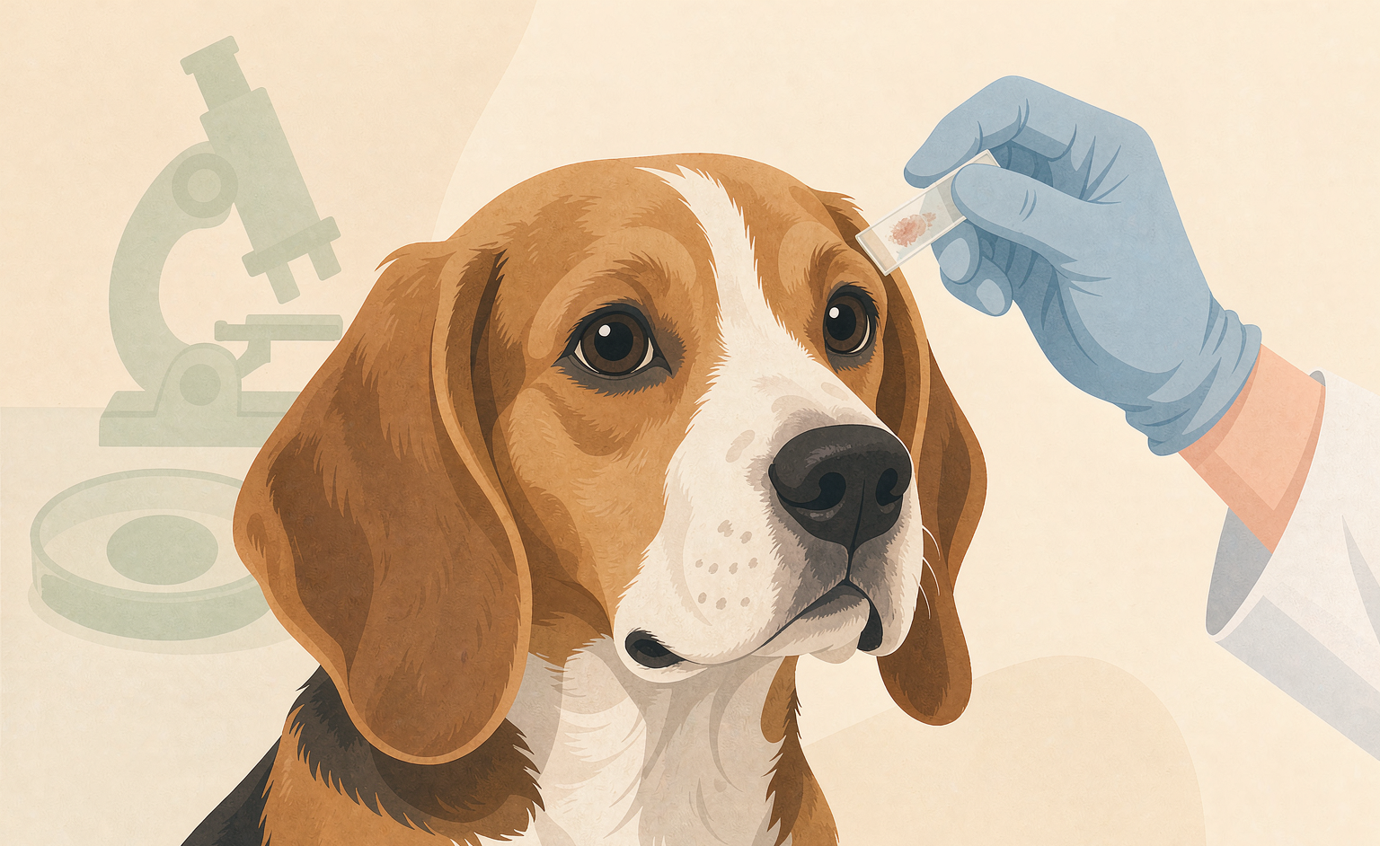
Canine yeast dermatitis is almost always caused by Malassezia pachydermatis, a fungus that normally lives on dog skin and overgrows when something else disrupts the skin barrier. It presents as greasy, thickened, reddened, itchy skin with a musty odor, most often in the ears, armpits, groin, paws, and skin folds. Diagnosis is by skin cytology, a quick in-clinic microscope test that confirms yeast organisms and rules out bacteria and mites, sometimes supported by culture or biopsy in chronic cases. Treatment uses topical antifungal therapy, with oral antifungals for extensive cases, but the underlying cause (allergies, hormonal disease, or anatomy) must be found and managed or the infection recurs. Antibiotics alone make yeast worse.
—
What Malassezia Actually Is and Why It Is Everywhere
Malassezia pachydermatis is a lipophilic yeast: it needs fat to grow, which is why it prefers the oily areas of canine skin. It is not an invader in the usual sense. It is a normal resident of the skin and ear canals of essentially every dog, present in small numbers in healthy animals without causing any problem at all.
The transition from normal resident to pathogen is driven by the skin environment. Several conditions make the skin friendlier to yeast:
Increased surface lipids. Greasy skin feeds Malassezia directly. Dogs with seborrheic skin, or those on high-fat diets, or with endocrine disease that alters sebum production, provide a richer food supply.
Increased moisture and occlusion. Skin folds, tight collars, and ear canals that do not ventilate well trap humidity. Yeast thrives in warm, damp, low-oxygen environments. This is why Bulldogs, Pugs, Shar-Peis, and other folded breeds are overrepresented.
Disrupted barrier and altered immunity. Allergic inflammation damages the skin barrier and changes the surface chemistry in ways that favor yeast. So does immunosuppression from corticosteroids, which is a common and underappreciated trigger.
Antibiotic exposure. Broad-spectrum antibiotics reduce the bacterial populations that normally compete with yeast for space and nutrients. A dog who develops a yeast infection after a course of antibiotics for something else is following a well-worn path.
The practical implication is that yeast overgrowth is a marker. It tells you the skin environment has changed, and the change is what deserves investigation.
Why It Gets Mistaken for Bacterial Infection
The overlap in presentation is genuine and it is the reason cytology matters.
Both bacterial pyoderma and yeast dermatitis can produce redness, itching, hair loss, thickened skin, and a bad smell. Both can affect the same locations. Owners and even experienced clinicians working from appearance alone cannot reliably separate them, and the error runs in one direction more often than the other: yeast gets treated as bacterial far more frequently than the reverse.
The consequences of that error are not neutral. Antibiotics do nothing to yeast. Meanwhile they reduce the competing bacterial flora, and the yeast population expands into the vacated niche. This is why the classic history of a chronic yeast case includes two or three courses of antibiotics with worsening skin. Getting the diagnosis right the first time prevents that cycle.
Two features push the suspicion toward yeast: the odor and the texture. Yeast dermatitis tends to smell musty, yeasty, or like corn chips, and the skin often feels greasy and looks thickened or elephant-like, with hyperpigmentation developing over time. Bacterial infections more often show papules, pustules, and epidermal collarettes, the circular scaly rings. These are tendencies, not rules, which is exactly why a microscope settles it.
The Tests That Actually Confirm It
Skin cytology is the primary test and the one that matters most. A vet presses a glass slide onto the affected skin, or takes a sample with a swab or tape, stains it, and looks under the microscope. Malassezia organisms appear as characteristic peanut or bottle-shaped yeasts, often in clusters. The test takes minutes, costs little, and gives a definitive answer on whether yeast are present and in what quantity. It also simultaneously reveals bacteria, and in many dogs both are present at once, which changes the treatment plan. Neutrophils on the slide indicate active inflammation and possible bacterial involvement.
Ear cytology is the same test applied to ear canal samples, and it is essential in any dog with otitis, because yeast otitis and bacterial otitis require different ear medications and the wrong one prolongs the problem.
Tape impression cytology is a variant used for dry or scaly lesions where pressing a slide does not collect enough material. Clear acetate tape is pressed to the skin, stained, and examined.
Skin scrapings look for mites, particularly Demodex, which can coexist with yeast and are a common reason a skin problem fails to resolve. Any chronic case deserves this.
Fungal culture is used less often, mainly for cases that do not respond to appropriate therapy or where a different fungal organism (Microsporum, Trichophyton for ringworm) is suspected. Malassezia culture has limited practical value because the organism is normal flora, so a positive culture alone does not prove disease; quantity and clinical signs matter.
Biopsy and histopathology is reserved for chronic, thickened, non-responsive cases, and it serves a different purpose: it identifies the deeper process, which might be a hypersensitivity, an endocrine-related change, or a condition like sebaceous adenitis.
Bloodwork for the underlying cause. Because yeast overgrowth so often signals an underlying problem, a chronic or recurrent case usually prompts screening for hypothyroidism and Cushing’s disease, plus a careful allergy history. This is not over-testing; the endocrine panel is often what finally explains a dog with a two-year history of skin problems.
Treatment: Topicals Do the Heavy Lifting
The treatment approach for yeast dermatitis has shifted firmly toward topical therapy, because topical antifungals reach the skin surface where the organism lives and avoid the systemic risks of oral drugs.
Medicated shampoos are the backbone for body lesions. Chlorhexidine combined with miconazole or ketoconazole is the standard formulation, used two to three times weekly with ten minutes of contact time before rinsing. The chlorhexidine addresses any concurrent bacteria, the azole addresses the yeast, and the combination is more effective than either alone in mixed infections. Selenium sulfide shampoos are an alternative with antifungal and oil-reducing effects.
Leave-on products extend the effect between baths: mousse, sprays, and wipes with chlorhexidine and an azole, particularly useful for skin folds, paws, and localized lesions. For fold dermatitis in wrinkled breeds, daily wiping and drying of the folds is not optional, it is the treatment.
Ear medications containing an azole, often combined with a steroid to reduce the intense inflammation, are the standard for yeast otitis. The steroid component is why ear cytology matters: using a steroid-containing ear medication in a dog with an intact eardrum and an undiagnosed problem can cause harm, and using an antibacterial-only ear drop on a yeast infection does nothing.
Oral antifungals such as ketoconazole, itraconazole, or fluconazole are used for extensive, deep, or refractory cases, and for dogs where topical therapy is impractical. They require liver monitoring in some cases, they interact with other drugs, and they are prescription-only. Ketoconazole in particular has meaningful drug interactions and endocrine effects, which is why the choice among azoles is a veterinary decision rather than a preference.
What does not work. Antibiotics, as covered. And corticosteroids, which reduce the itch dramatically and are often what owners ask for, suppress local immunity and feed the underlying problem. A dog on steroids for itchy skin who develops a worsening yeast infection is following the expected course. Steroids have a legitimate role in managing the allergic component, but only with antifungal cover and veterinary supervision.
The Underlying Cause Is the Actual Case
This is the part that determines long-term outcome, and it is where most of the effort belongs once the yeast is under control.
In a large proportion of chronic canine yeast dermatitis cases, the driver is allergic skin disease, either environmental atopy, food allergy, or flea allergy. The mechanism is straightforward: allergic inflammation damages the barrier and alters surface lipids, yeast overgrow in the disrupted environment, and treating the yeast without treating the allergy produces a dog who improves for a few weeks and relapses. Our dog itching guide walks through the allergy workup, and the skin problems overview covers the broader diagnostic ladder.
Endocrine disease is the second major driver. Hypothyroidism slows skin turnover and alters sebum, and Cushing’s disease thins the skin and suppresses immunity. Both produce recurrent yeast and bacterial infections, and both are treatable. A middle-aged or older dog with a first-time chronic yeast problem is a reasonable candidate for thyroid and adrenal screening.
Anatomy is the third, and it is the one most amenable to management rather than cure. Folded skin, narrow ear canals, and hairy ear canals will always be favorable environments. These dogs need a maintenance routine rather than a one-time cure: regular fold cleaning and drying, hair plucking in the ear canals where appropriate, and ongoing medicated bathing at a reduced frequency.
The maintenance plan for a dog with recurrent yeast typically looks like this: identify and manage the underlying cause, use medicated shampoo every one to two weeks indefinitely rather than stopping when the skin clears, keep folds and ears dry, and recheck cytology periodically rather than judging by appearance alone, because yeast can be present in numbers that matter before the skin looks bad again.
Where Daily Support Fits
Dogs with chronic yeast and allergy problems have skin that needs ongoing support, and there is a nutritional layer to that which is worth understanding separately from treatment.
The skin barrier is built from dietary fat, omega-3 fatty acids, zinc, and B vitamins including biotin, and a barrier running short on these heals slowly and flares easily. Omega-3 fatty acids in particular have a consistent evidence base in reducing the inflammatory and itching component of allergic skin disease, which matters here because scratching and self-trauma are what convert a mild yeast overgrowth into a spreading lesion. Our omega-3 soft chews are built around EPA and DHA with a skin-support base, and our yeast balance formula combines ingredients chosen for dogs dealing with recurrent yeast and skin balance in a daily chew. Both are daily nutritional support, not a treatment for yeast dermatitis or any active infection, which needs cytology, antifungal therapy, and management of the underlying cause. Used as the maintenance layer under a vet’s plan, they support the barrier that keeps the problem from coming back.
Frequently Asked Questions
How do I know if my dog has a yeast infection or a bacterial infection?
You cannot reliably tell by looking, which is why skin cytology exists. Yeast dermatitis tends to smell musty or like corn chips and the skin feels greasy and thickened, while bacterial infections more often show pimples and circular scaly rings. Both can appear in the same places and both itch. A vet pressing a slide onto the skin and examining it under a microscope settles the question in minutes and often reveals both organisms at once.
What does a dog yeast infection look like?
Greasy, reddened, thickened skin with a musty odor, often with hyperpigmentation developing over time, in the ears, armpits, groin, paws, and skin folds. Itching is usually intense, and the dog may lick, chew, or scoot. Chronic cases develop an elephant-like thickening of the skin called lichenification. The appearance overlaps heavily with bacterial pyoderma and with allergic dermatitis.
Why did my dog get a yeast infection after antibiotics?
Because antibiotics reduce the bacterial populations that normally compete with yeast for space and nutrients on the skin. With the competition removed, *Malassezia* expands into the vacated niche. This is one of the most common sequences in veterinary dermatology, and it is a strong argument for confirming a diagnosis with cytology before starting antibiotics.
Can I treat my dog’s yeast infection at home without a vet?
Mild, localized cases sometimes respond to medicated chlorhexidine and miconazole shampoo used correctly, with proper contact time. But home treatment without a diagnosis risks treating the wrong organism, and it will not address the underlying allergy or endocrine problem that caused the overgrowth. A dog with spreading, painful, or recurrent lesions needs cytology and a plan.
How long does it take to clear a dog yeast infection?
Visible improvement usually comes within one to two weeks of appropriate topical therapy, and full resolution of thickened, pigmented skin can take one to three months. The more important number is the recurrence rate: without identifying and managing the underlying cause, most dogs relapse within weeks to months, which is why treatment does not end when the skin looks better.
How often should I bathe my dog with yeast dermatitis?
Two to three times weekly during active treatment, with ten minutes of contact time before rinsing, then tapering to once every one to two weeks for long-term maintenance in dogs prone to recurrence. Bathing too infrequently is a common reason treatment fails, and rinsing off the shampoo too quickly is another.
Is dog yeast infection contagious to other pets or humans?
*Malassezia pachydermatis* is normal skin flora on dogs and is not considered contagious in the usual sense. Other pets in the household are not at meaningful risk, and human infection from a dog is rare and generally limited to people who are significantly immunocompromised. The dog’s own compromised skin barrier is the real issue.
Bottom Line
Skin yeast infection in dogs is one of the most frequently misdiagnosed problems in veterinary dermatology, and the misdiagnosis has a cost: antibiotics do nothing to yeast and actively clear the way for it to spread. The fix is cheap and fast, a skin cytology that takes minutes and shows exactly what is on the skin, including the mixed infections that appearance alone would miss. Once confirmed, topical antifungal therapy does most of the work, with oral drugs reserved for extensive cases. But the part that actually decides the outcome is upstream: yeast overgrowth is a signal that the skin environment has changed, and whether that change is allergy, hypothyroidism, Cushing’s, or simply the geometry of a folded face determines whether this is a two-week episode or a recurring feature of the dog’s life. Find the signal, not just the organism.
Disclaimer
This article is for general education only and is not veterinary advice. Yeast dermatitis can resemble other skin diseases including bacterial infections, ringworm, and demodectic mange, and only cytology or culture can distinguish them. Prescription antifungal medications require veterinary supervision and, in some cases, liver monitoring. If your dog has spreading, painful, or non-healing skin lesions, or is systemically unwell, contact a veterinarian promptly. Dietary supplements are not intended to diagnose, treat, cure, or prevent any disease, and you should consult your veterinarian before starting any supplement.
References
- Merck Veterinary Manual. “Malassezia Dermatitis in Dogs.”
- VCA Animal Hospitals. “Yeast Dermatitis in Dogs” and “Malassezia Otitis.”
- Cornell University College of Veterinary Medicine. “Cutaneous Malassezia Infections in Dogs.”
- Miller WH, Griffin CE, Campbell KL. “Muller and Kirk’s Small Animal Dermatology.” Elsevier.
- Bond R, et al. “Malassezia pachydermatis: a review.” Medical Mycology.